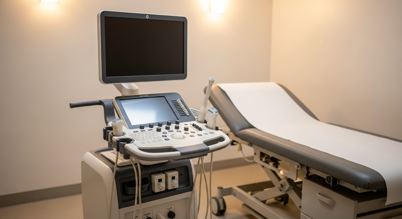
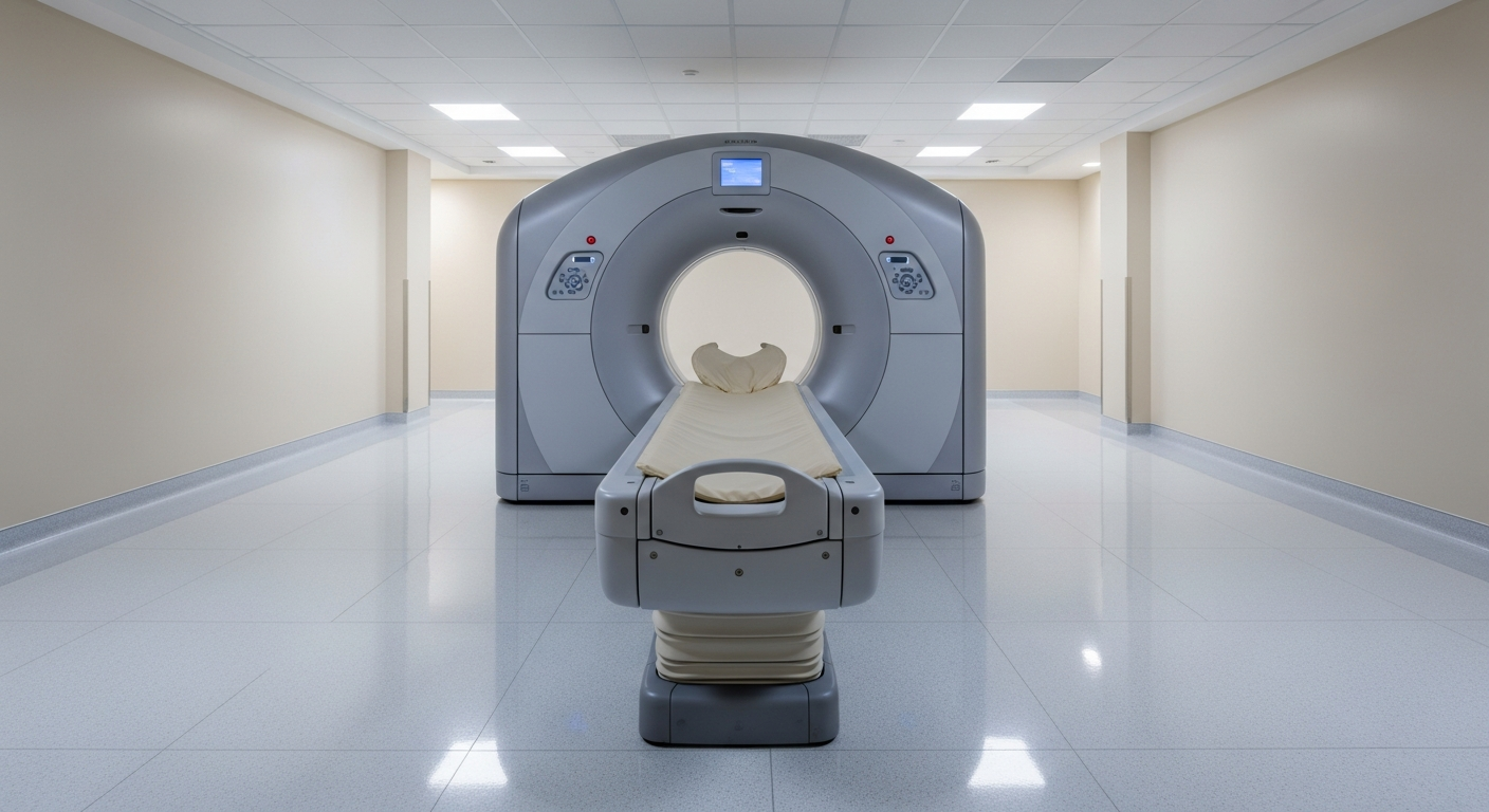
X-Ray vs MRI — When to Choose Each
Compare X-ray and MRI — what each shows best, radiation, scan time, cost, and how clinicians decide which imaging to order first.

X-ray and MRI sit at opposite ends of the imaging spectrum: X-ray is fast, cheap, and shows bone; MRI is detailed, longer, and shows soft tissue. Most clinical work-ups use them in sequence rather than one in place of the other.
TL;DR comparison
| Feature | X-Ray | MRI |
|---|---|---|
| How the image is made | A single X-ray beam through tissue | Magnetic field + radio waves |
| Ionizing radiation | Yes — a very low dose | None |
| Typical scan time | A few minutes | 20–45 minutes |
| Best at showing | Bone, fractures, lungs, joint alignment | Soft tissue, ligaments, brain, spine, marrow |
| Real-time / weight-bearing? | Often yes (standing studies) | No (supine, in the bore) |
| Sedation needs | Almost never | Sometimes for severe anxiety |
| Cost | Lowest of all modalities | Higher |
| Available at Precise | Yes — every imaging site | Yes — most sites |
How each modality works
X-ray sends a single beam of low-dose ionizing radiation through the body. Dense tissues like bone block more of the beam and appear bright on the image; soft tissue blocks less and appears darker. The result is a 2D projection that's exceptionally good for bones, alignment, and basic chest pathology.
MRI uses a strong magnet and radio-frequency pulses to map how water and fat behave in soft tissues. It produces multi-planar cross-sectional images with no radiation exposure — and shows ligaments, tendons, cartilage, nerves, brain, and bone marrow in detail no X-ray can match.
When X-ray is preferred
- Suspected acute bone fracture
- Joint dislocation or alignment evaluation
- Routine chest X-ray for cough, pneumonia, or pulmonary nodule screening
- Pre-operative orthopedic planning (weight-bearing alignment)
- Foreign body localization
- Quick follow-up after fracture reduction or hardware placement
When MRI is preferred
- Soft-tissue injury (ligament, tendon, meniscus, cartilage)
- Disc herniation, nerve impingement, or spinal cord pathology
- Brain conditions (stroke follow-up, MS, tumors, seizures)
- Bone marrow edema, occult fractures not visible on X-ray
- Joint internal derangement when X-rays are normal
- Soft-tissue masses needing tissue characterization
Safety, speed, and cost differences
Radiation. X-ray exposes you to a very small dose of ionizing radiation — typically 0.001–0.1 mSv for an extremity or chest film. MRI uses none. For most adults, the X-ray dose is negligible; for pregnant patients or children, the radiologist may suggest alternatives when possible.
Speed. X-ray takes minutes from check-in to leaving. MRI takes 20–45 minutes on the table, plus check-in and changing.
Cost. X-ray is the least expensive of the major imaging studies in both insurance and self-pay scenarios. MRI is among the more expensive.
Comfort and access. X-ray is open and quick — most patients are in and out in under 15 minutes. MRI requires lying still in an enclosed (or open) magnet for the duration of the scan. Wide-bore 3T or Open MRI at Precise helps if you're claustrophobic.
Implants and contraindications. X-ray has essentially none beyond pregnancy precautions. MRI requires safety screening for pacemakers, neurostimulators, certain implants, and metal fragments.
Order of operations. It's common to start with X-ray and add MRI only if X-ray is inconclusive or soft-tissue injury is suspected. For acute trauma the typical flow is X-ray → MRI if persistent symptoms or normal X-ray.
How to decide with your doctor
The two tests answer different questions and are often complementary. Your physician will consider:
- What's most likely the cause of your symptoms. Bone-and-alignment questions → X-ray first. Soft-tissue, neurologic, or marrow questions → MRI.
- Cost and access. X-ray is faster and cheaper and is often the right first step.
- Radiation history. When prior imaging exists, MRI may avoid an additional X-ray dose.
- Specifics of the injury. For ongoing back pain after a normal X-ray, MRI is the next step. See MRI vs X-ray for back pain for that decision in depth.
To schedule, see X-ray services or MRI services, or call 800-558-2223 for help.
Frequently asked questions
Should I get an X-ray or MRI first for joint pain?+
For an acute injury where a fracture is suspected, X-ray is usually the first study. If X-ray is normal and pain persists, or if soft-tissue injury (ligament, tendon, meniscus) is suspected, MRI is typically next.
Does an MRI show what an X-ray can't?+
Yes — MRI shows ligaments, tendons, cartilage, the spinal cord and nerves, brain tissue, and bone marrow, none of which X-ray can directly visualize. X-ray, in turn, is fast and excellent for fractures, alignment, and lung pathology.
Is one of these tests more accurate?+
Neither is universally more accurate — they answer different questions. The right test depends on whether the suspected problem is bony or soft-tissue.
How much radiation is in an X-ray compared to MRI?+
MRI uses no ionizing radiation. A typical extremity or chest X-ray exposes you to about 0.001–0.1 mSv — a very small dose for diagnostic exams.
Can a single visit cover both X-ray and MRI?+
At many Precise locations, yes — both modalities are on site. Schedulers can sometimes coordinate same-day imaging when both are ordered.