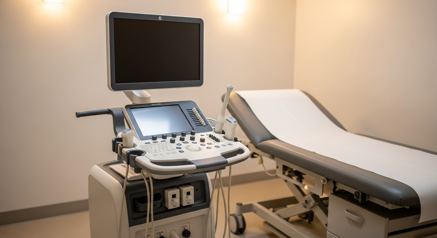
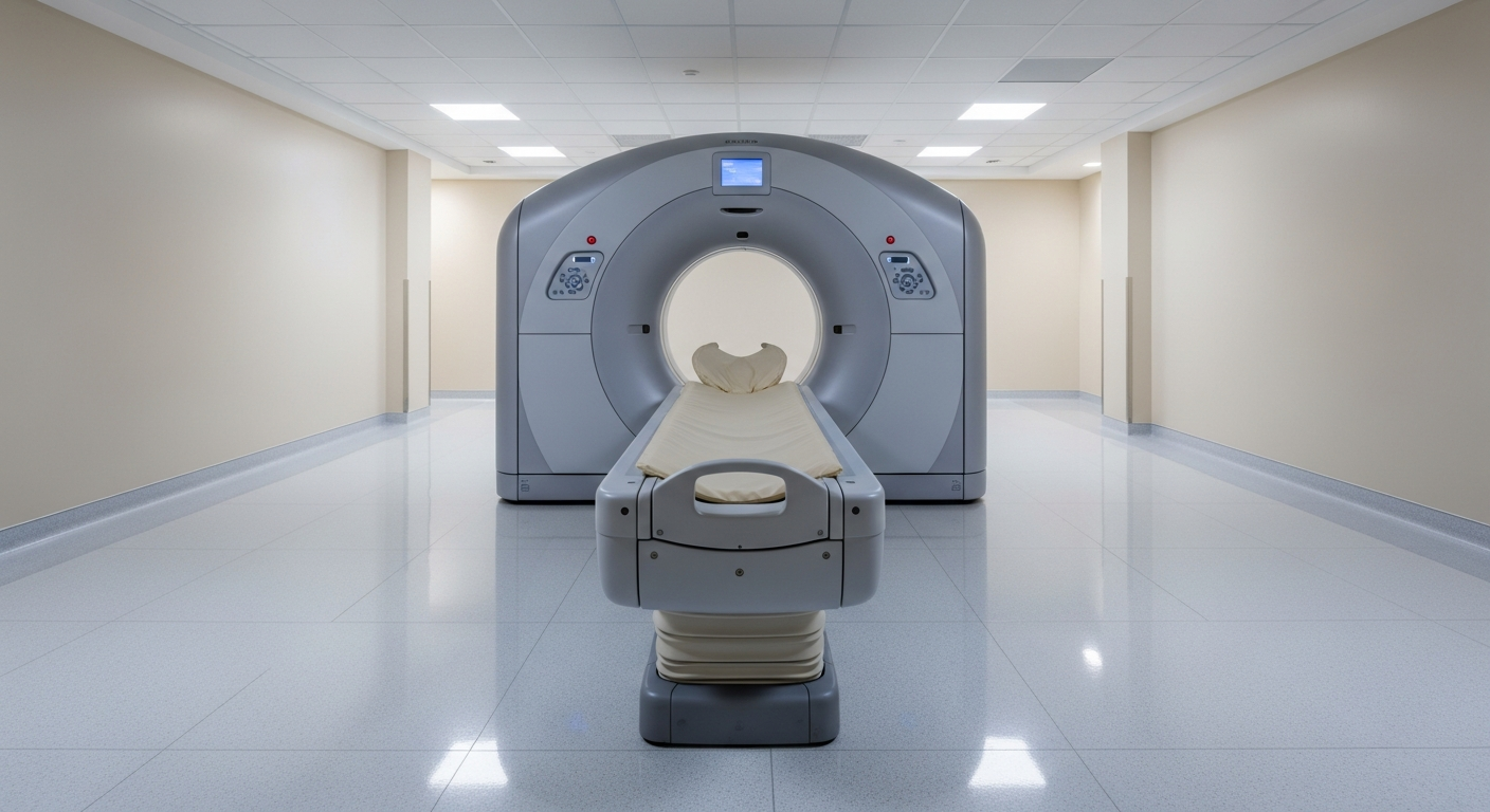
MRI vs X-Ray for Back Pain — Which Should You Get First?
For most adults with new back pain, neither imaging test is needed at first. When it is, X-ray and MRI answer very different questions. Here's how to decide.

Most new low-back pain in adults gets better within 4–6 weeks with conservative care, and current evidence-based guidelines (American College of Physicians, ACR Appropriateness Criteria) recommend no imaging initially in the absence of red-flag symptoms. When imaging is appropriate, X-ray and MRI answer very different questions.
TL;DR comparison
| Feature | X-Ray of spine | MRI of spine |
|---|---|---|
| Best at showing | Alignment, vertebral fractures, degenerative bony changes | Disc herniation, nerve compression, spinal cord, marrow |
| Radiation | Low dose | None |
| Scan time | A few minutes | 25–40 minutes |
| Weight-bearing? | Yes (standing films possible) | No (supine in the bore) |
| When ordered first | Acute trauma, suspected fracture or instability | Persistent radiculopathy, suspected disc/nerve cause, red flags |
| Cost | Lowest of all spine imaging | Higher |
| Available at Precise | Yes | Yes — including 3T and Open MRI |
When imaging is and isn't appropriate
For most adults with new low-back pain:
- No red-flag symptoms + pain less than 4–6 weeks: imaging is generally not recommended. Studies show early imaging does not improve outcomes and can lead to unnecessary procedures.
- Conservative care first: relative rest, NSAIDs as appropriate, physical therapy, gradual return to activity.
Red-flag symptoms that warrant imaging now (talk to your physician):
- New bowel or bladder incontinence, or saddle anesthesia (possible cauda equina — urgent)
- Major trauma, especially in patients over 50 or with osteoporosis
- Progressive neurologic deficit (weakness, foot drop, worsening numbness)
- Fever, unexplained weight loss, or history of cancer
- IV drug use, immunosuppression, or suspected infection
- Pain that wakes you at night and is not relieved by position change
- Pain after a fall in an older adult or known osteoporotic patient
When X-ray is the right first imaging
- Suspected acute vertebral compression fracture after a fall
- Trauma in a patient old enough that DEXA / bone-density status matters
- Suspected spinal instability or spondylolisthesis (flexion-extension views)
- Pre-procedure or pre-operative planning
- Scoliosis evaluation (standing full-length films)
- Hardware follow-up after spine surgery
When MRI is the right first imaging
- Persistent radiculopathy (sciatica, leg numbness/weakness) after 4–6 weeks
- Any red-flag symptom suggesting cord, nerve root, infection, or tumor
- Failed conservative care and surgical consultation is being considered
- Suspected disc herniation, spinal stenosis, or annular tear
- Inflammatory back pain (suspected sacroiliitis or ankylosing spondylitis)
- Suspected vertebral osteomyelitis or epidural abscess (urgent)
- Suspected cord lesion (myelopathy, MS)
Safety, speed, and cost differences
Radiation. Lumbar X-ray exposes you to roughly 0.5–1.5 mSv per study. MRI is radiation-free.
Speed. X-ray takes minutes. MRI requires 25–40 minutes on the table plus check-in.
Detail. MRI is the only test that shows the spinal cord, nerve roots, and disc material — the structures that actually cause radiating pain. X-ray cannot show these directly.
Cost. X-ray is the least expensive spine imaging. MRI is more expensive but is the test that answers most surgical-planning questions.
Order of operations. A common pattern is: conservative care → X-ray if trauma or fracture suspected → MRI if radiculopathy persists or red flags emerge.
How to decide with your doctor
- Mention every red-flag symptom clearly when you see your physician. They change the urgency and modality.
- Avoid jumping straight to MRI for uncomplicated, recent-onset back pain — most patients get better with conservative care.
- Ask whether weight-bearing films are needed for suspected instability or scoliosis.
- Bring prior imaging if you have it. The radiologist will compare side-by-side.
- Schedule MRI on a 3T or Open MRI if you've struggled with closed-bore scanners — see 3T vs 1.5T MRI and Open vs Closed MRI.
For broader context on the two modalities, see X-ray vs MRI. To schedule, see MRI services or X-ray services, or call 800-558-2223.
Frequently asked questions
Do I need imaging for new back pain?+
For most adults with new low-back pain and no red-flag symptoms, current evidence-based guidelines recommend no imaging in the first 4–6 weeks. Most patients improve with conservative care. Imaging is appropriate when red flags are present or symptoms persist or worsen.
What are red-flag symptoms that mean I need imaging now?+
New bowel or bladder incontinence, saddle anesthesia, major trauma, progressive neurologic deficit, fever with back pain, unexplained weight loss, history of cancer, IV drug use or immunosuppression, and pain that wakes you at night.
Should I just get an MRI to be sure?+
Not as a first step in uncomplicated low-back pain. Early MRI without indication does not improve outcomes and can lead to incidental findings that drive unnecessary procedures. Discuss the right timing with your physician.
What does an X-ray of the back show?+
Bony alignment, vertebral compression fractures, degenerative bony changes, spondylolisthesis (especially with flexion-extension views), scoliosis, and post-surgical hardware. X-ray does not show discs, nerve roots, or the spinal cord directly.
What does an MRI of the back show?+
Discs and disc herniation, spinal cord, nerve roots, marrow edema, inflammation, infection, tumor, and soft-tissue detail X-ray cannot show — making MRI the right test for radiculopathy, suspected nerve compression, or red-flag work-ups.
How long should I wait before getting an MRI for back pain?+
If you have no red flags, most clinicians recommend conservative care for at least 4–6 weeks before considering MRI. If you develop red-flag symptoms or persistent radiculopathy, talk to your physician sooner.