CT vs Ultrasound — When Each Test Is the Right Call
CT and ultrasound both look inside the body, but one uses radiation and cross-sectional detail while the other is radiation-free and real-time. A plain guide to how physicians choose.

CT and ultrasound are both cornerstone tools for looking inside the body, but they work on completely different physics and shine in different situations. One delivers detailed cross-sectional slices using X-rays; the other gives a live, radiation-free view using sound. Knowing how they differ helps explain why your doctor chose one over the other.
TL;DR comparison
| Feature | CT | Ultrasound |
|---|---|---|
| How the image is made | Rotating X-ray beams reconstructed into 3-D slices | High-frequency sound waves reflected back by tissues |
| Ionizing radiation | Yes — moderate dose (2–10 mSv typical) | None |
| Best at showing | Lungs, bone detail, bleeding, complex trauma, deep organs | Soft tissue, fluid, blood flow, gallbladder, pelvic and abdominal organs |
| Real-time imaging? | No — a captured study | Yes — dynamic, live view |
| Contrast agent | Often (IV iodinated contrast) | Usually none (Doppler shows flow without contrast) |
| Operator dependence | Low — standardized protocols | Higher — depends on sonographer skill |
| Typical scan time | 30 seconds – 5 minutes | 20–45 minutes |
| Typical out-of-pocket (cash) | Higher | Low |
| Available at Precise | Yes — multidetector CT | Yes |
How each modality works
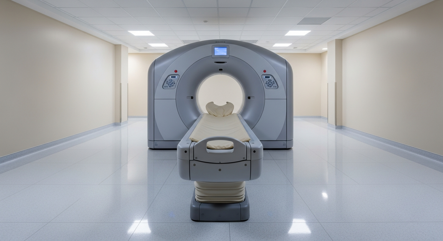
A CT (computed tomography) scanner rotates an X-ray tube around the body while a ring of detectors measures how much radiation passes through each tissue. A computer reconstructs hundreds of thin slices into detailed cross-sectional and 3-D images, letting the radiologist view organs, vessels, bone, and soft tissue from any angle.
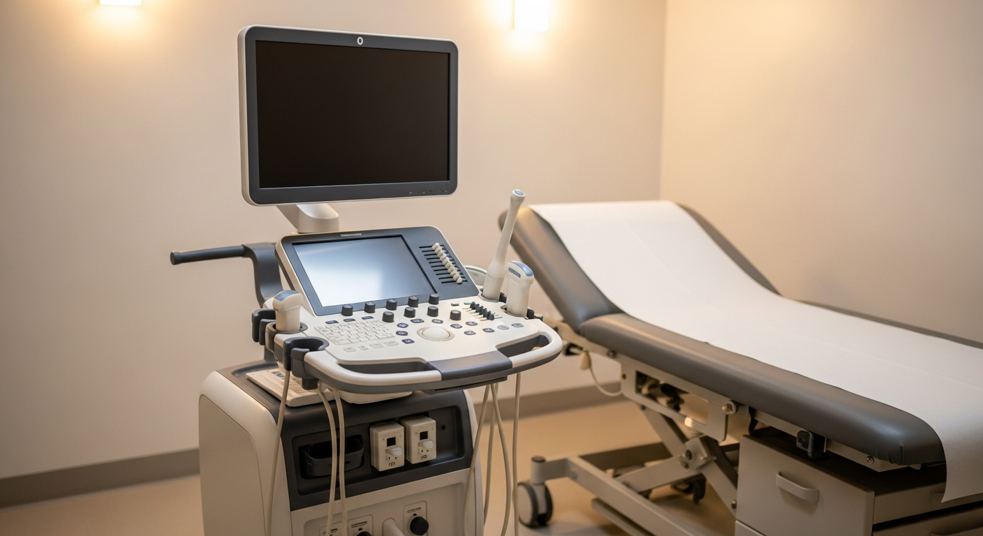
Ultrasound sends pulses of high-frequency sound (2–18 MHz) into the body through a handheld transducer. The waves bounce off tissue interfaces and the returning echoes are reconstructed into a live, real-time image. No radiation is involved. Doppler ultrasound adds color-coded maps of blood velocity and flow direction.
When CT is the right test
- Acute trauma — rapid, whole-region assessment of organs, bleeding, and complex fractures
- Suspected pulmonary embolism, aortic dissection, or aneurysm (CT angiography)
- Lung disease — nodules, pneumonia detail, interstitial disease
- Cancer staging, surveillance, and detection of metastases
- Kidney stones and many causes of acute abdominal pain in larger patients
- Complex or spinal fractures where 3-D bone detail matters
When ultrasound is the right test
- Gallbladder and biliary evaluation — gallstones, cholecystitis
- Pelvic pain or abnormal uterine bleeding — uterus and ovary assessment
- Obstetric imaging — pregnancy dating, fetal anatomy, placenta location
- Palpable lump or mass in the neck, breast, thyroid, or soft tissue
- Vascular assessment — deep vein thrombosis (DVT), carotid stenosis, peripheral arteries
- Scrotal pain or testicular mass
- Procedural guidance — needle biopsies, fluid drainage, IV placement
- First-line abdominal imaging in children and pregnant patients (no radiation)
Radiation and safety
CT uses ionizing radiation — typically 2–10 mSv depending on the body region and protocol. For a single diagnostic exam the absolute risk is small, but physicians weigh dose against benefit, especially for children, pregnant patients, and anyone needing repeated imaging.
Ultrasound uses no ionizing radiation at all, which is why it is the preferred first test in pregnancy, in children, and whenever a clinical question can be answered without radiation. There are no known harmful effects from diagnostic ultrasound at standard clinical settings.
Limitations of each modality
CT limitations:
- Uses ionizing radiation, so repeat imaging adds cumulative dose
- IV iodinated contrast requires kidney-function screening and carries a small reaction risk
- Soft-tissue contrast is good but not as detailed as MRI for some structures
Ultrasound limitations:
- Sound waves do not penetrate bone or gas — deep structures and obese patients can be hard to see
- Bowel gas degrades abdominal assessment significantly
- Image quality and completeness depend on the sonographer's skill and patient body habitus
- Cannot image the lungs, brain, or spinal cord (bone and air block the sound)
How to decide with your doctor
- Is the question about the gallbladder, pelvis, a vessel, or a lump? → Ultrasound is usually first.
- Is it acute trauma, the lungs, or complex bone? → CT is usually first.
- Is the patient pregnant or a young child? → Ultrasound first (no radiation); CT only when clearly indicated.
- Does the problem need real-time or dynamic evaluation, like blood flow or compressibility? → Ultrasound shows movement and flow that CT cannot.
- Normal ultrasound but symptoms persist, or deeper detail needed? → CT (or MRI) is typically next.
If you have a referral and want help choosing or scheduling the right first test, call us at 800-558-2223 or visit CT services or ultrasound services.
Frequently asked questions
Is CT or ultrasound better?+
Neither is universally better — they answer different questions. Ultrasound is excellent for the gallbladder, pelvis, blood vessels, and superficial masses; CT is superior for the lungs, acute trauma, complex bone, and deep structures. Your physician picks based on the clinical question.
Why would my doctor order ultrasound before CT?+
Ultrasound is fast, inexpensive, uses no radiation, and gives real-time information. For many initial work-ups — gallbladder pain, a palpable lump, a vascular concern, or pelvic symptoms — it answers the question immediately without exposing you to radiation.
Does CT use radiation and ultrasound doesn't?+
Yes. CT uses ionizing radiation — typically 2–10 mSv depending on the body region and protocol. Ultrasound uses high-frequency sound waves and emits no radiation at all, which is why it is the first choice in pregnancy and in children.
Can ultrasound see everything a CT can?+
No. Sound waves cannot penetrate air or bone, so ultrasound cannot evaluate the lungs, the inside of bone, or the brain and spinal cord. Bowel gas and body habitus can also limit deep abdominal views. CT remains the right test for those situations.
Is ultrasound cheaper than CT?+
Yes, in most insurance and self-pay scenarios. Ultrasound is one of the most affordable imaging tests, while CT costs more and uses more radiation. Precise publishes transparent self-pay rates and provides a written estimate before the exam.
Can I have both an ultrasound and a CT for the same problem?+
Yes, and it's common. For example, an abdominal ultrasound may be the first study for right-upper-quadrant pain, and a CT may follow when the picture is unclear or deeper detail is needed. Your physician will sequence the studies to answer one question at a time.