Rotator Cuff Imaging
How X-ray, MRI, and MR arthrography fit into the work-up of suspected rotator cuff disease.

The rotator cuff is a group of four muscles and their tendons that stabilize the shoulder and produce most of its rotational movement. Tears and tendinopathy are common, particularly after age 40 and in athletes who perform overhead activities. This guide explains the typical imaging sequence and what each study contributes.
This article is educational. It does not replace medical advice. Treatment decisions belong to your orthopedic surgeon or sports-medicine physician.
Step one: history and examination
The diagnosis usually starts with shoulder pain, weakness with specific movements, or a clear injury event. Specific provocative tests (Jobe's, drop-arm, lift-off, Hawkins-Kennedy) help localize the problem to the rotator cuff and differentiate it from impingement, biceps tendon pathology, or labral injury.
Step two: X-ray
Plain radiographs are usually the first imaging step. X-ray shows the bony anatomy of the shoulder, including the position of the humeral head, the acromion shape, calcific tendon deposits, arthritis, and any subtle bony avulsions. Walk-in X-ray is available at most Precise locations.
Step three: MRI
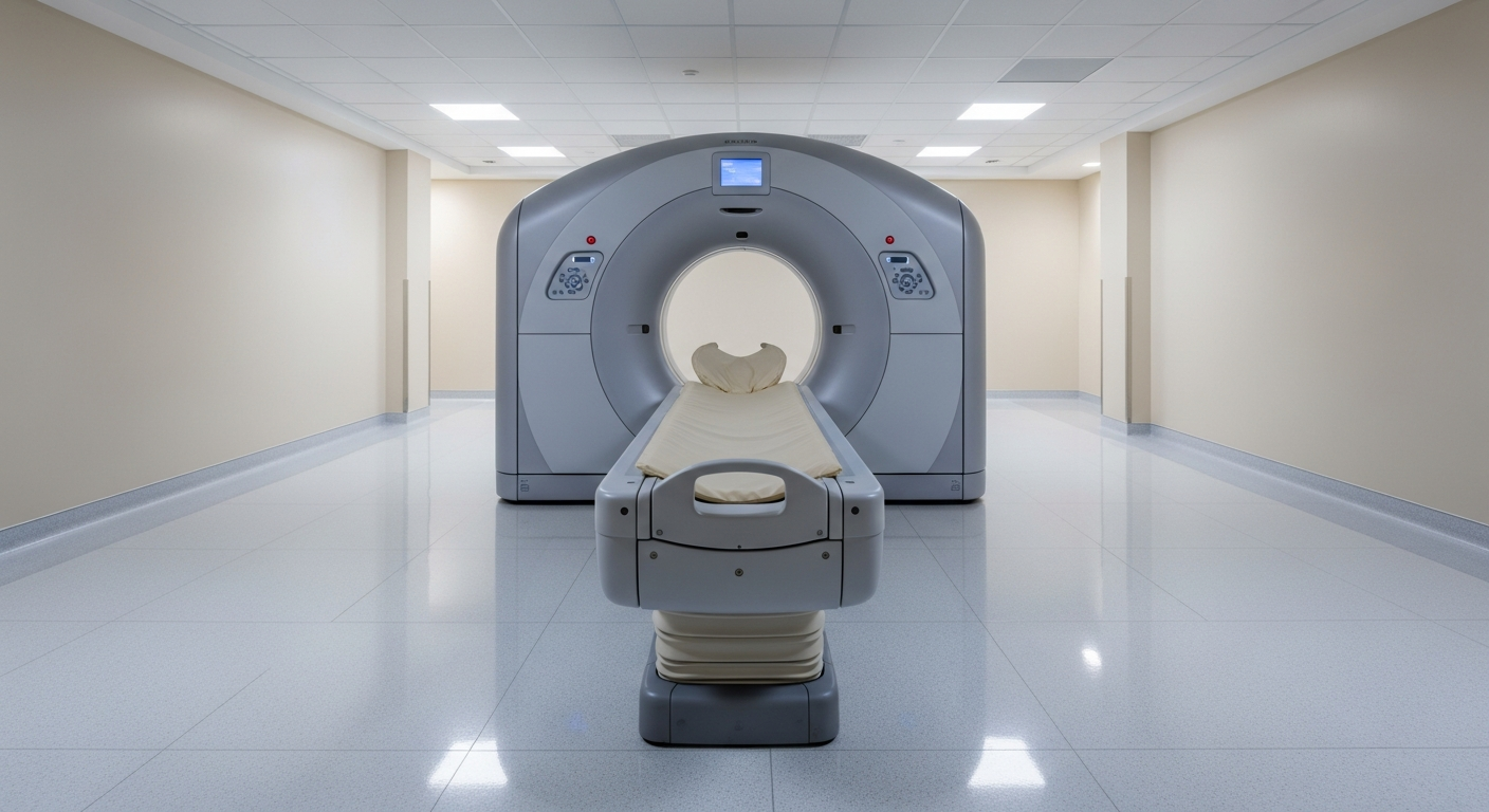
MRI is the workhorse for soft-tissue evaluation of the rotator cuff. It shows the tendons, the muscle bellies, the subacromial bursa, the labrum, the biceps anchor, and the surrounding bone. The radiologist describes whether a tear is partial- or full-thickness, the size and location, any retraction, muscle atrophy, and fatty infiltration. Each of those features matters to a surgeon planning a repair.
A standard shoulder MRI at Precise Imaging takes 20–35 minutes.
Step four: MR arthrography (when needed)
MR arthrography adds an intra-articular contrast injection — typically a gadolinium-based mixture — performed under fluoroscopic or ultrasound guidance immediately before the MRI. The contrast distends the joint capsule and dramatically improves visualization of:
- Labral tears (especially SLAP lesions and Bankart lesions).
- Articular-sided partial rotator cuff tears that can be subtle on standard MRI.
- Selected ligament injuries.
Not every shoulder MRI needs arthrography. Your orthopedic surgeon or sports-medicine physician decides whether to add it based on the clinical question. Precise Imaging performs MR arthrography at select locations.
What the report tells the surgeon
A useful rotator cuff MRI report describes:
- The specific tendon involved (supraspinatus, infraspinatus, subscapularis, teres minor).
- Partial- vs. full-thickness tear.
- Tear size in two dimensions.
- Tendon retraction.
- Muscle atrophy and fatty infiltration (Goutallier grading).
- Associated findings (labral tear, biceps pathology, AC joint arthritis, subacromial impingement features).
These features predict whether a tear is repairable, how it should be repaired, and the likely outcome.
What we offer for shoulder work-ups
- Shoulder MRI (1.5T and 3T).
- MR arthrography for selected labral or rotator cuff questions.
- Fluoroscopic arthrogram for image-guided intra-articular contrast injection.
- Walk-in X-ray at most locations.
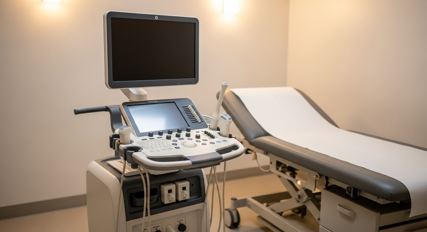
We do not currently advertise musculoskeletal ultrasound. When MSK ultrasound is needed, please refer to a center that offers it.
Frequently asked questions
Will I need contrast for my shoulder MRI? Most initial shoulder MRIs do not require intravenous contrast. MR arthrography uses intra-articular contrast for selected labral or partial-tear questions and is ordered by your surgeon when the clinical question warrants it.
How long is the appointment? Plan for 20 to 35 minutes of scan time for a standard shoulder MRI, plus 15 to 20 minutes for check-in and positioning. MR arthrography adds 20 to 30 minutes for the fluoroscopic or ultrasound-guided injection before the MRI begins.
Is the arthrogram injection painful? Most patients describe brief pressure during the injection and minimal pain. The radiologist performing the injection uses local anesthetic to numb the skin and joint capsule. Mild post-injection soreness for a day or two is common and resolves on its own.
Do I need to do anything special after MR arthrography? Rest the shoulder for the remainder of the day, avoid heavy lifting for 24 hours, and use ice if you feel soreness. Most patients return to normal activity the next day. Significant pain, swelling, fever, or redness should prompt a call to your provider.
Can I have a shoulder MRI with a rotator cuff repair anchor or plate? Yes. Modern shoulder hardware is MRI-conditional and metal-aware protocols reduce artifact substantially. Tell our scheduling team about prior surgery and hardware so the technologist can choose the appropriate protocol.
Will the surgeon get my report quickly? Final reports are typically signed within 24 to 48 hours and sent directly to your surgeon's office. Same-day or STAT turnaround is available on request.
Related Precise services
Shoulder work-ups draw on three Precise services: walk-in X-ray at most locations for initial bony evaluation, our MRI program (1.5T and 3T) for definitive soft-tissue imaging, and MR/fluoroscopic arthrogram for labral and selected partial-tear questions. The orthopedic and sports imaging hub connects these services with the supporting articles in this series.
Disclaimer
This article is for general patient education and does not replace medical advice from your physician. Rotator cuff management depends on individual factors and belongs to your treating orthopedic team.
For more, see ACL injury imaging pathway, return-to-play imaging, post-op imaging basics, or visit the orthopedic and sports imaging hub.