Post-Concussion Imaging — What MRI Can and Can't Tell You
A patient-friendly look at when imaging is appropriate after a concussion, and what it can and cannot show.

Concussions are a form of mild traumatic brain injury. They are common in sports, motor-vehicle collisions, and falls. Most concussions are diagnosed clinically — without imaging — because routine imaging is normal in the majority of patients. This guide explains when imaging is appropriate, what MRI can and cannot show after a concussion, and how to think about follow-up.
This article is educational. It does not replace medical advice. If you have a head injury with loss of consciousness, persistent vomiting, worsening headache, weakness, confusion, or any other concerning symptom, seek emergency care immediately.
What concussion is — and isn't
A concussion is a functional brain injury, not primarily a structural one. The symptoms — headache, dizziness, brain fog, sensitivity to light or noise, sleep disturbance, mood change — reflect disruption of brain network function rather than visible damage on standard imaging. That is why a "normal" MRI does not rule out a concussion.
When acute imaging is appropriate
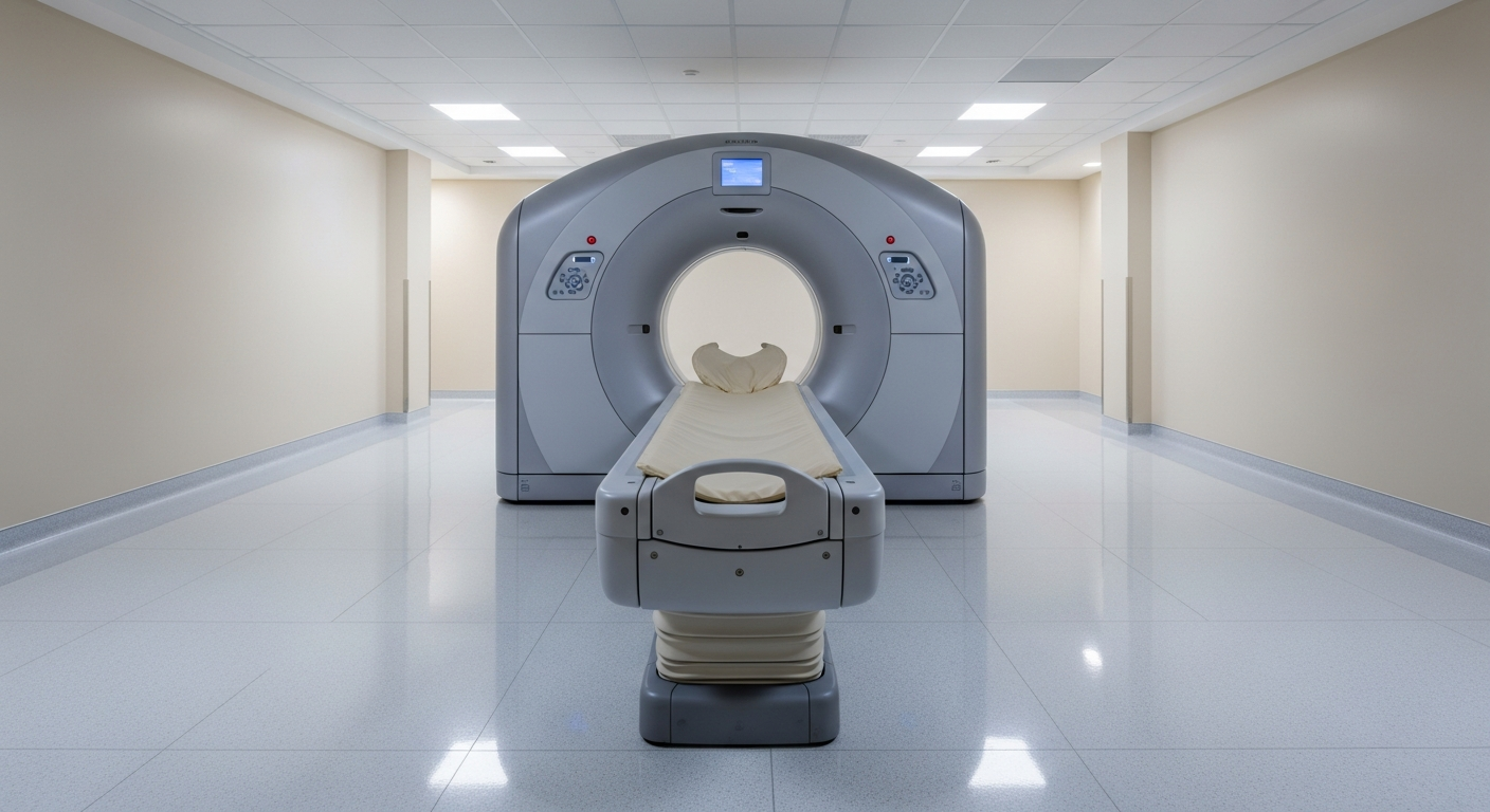
In the acute setting, CT — not MRI — is the first imaging study used to rule out a more serious injury such as a skull fracture, brain bleed, or contusion. Validated clinical decision rules (such as the Canadian CT Head Rule and PECARN criteria for children) help emergency physicians decide which patients need a CT after a head injury. Concerning features include loss of consciousness, persistent vomiting, severe or worsening headache, focal neurological signs, suspected skull fracture, or anticoagulant use.
When sub-acute or chronic MRI is appropriate
In the days to weeks after a concussion, MRI is sometimes ordered when:
- Symptoms persist longer than expected.
- New or progressive neurological symptoms appear.
- The clinical picture suggests something other than a simple concussion (for example, an underlying lesion).
- A patient with prior brain injury is being re-evaluated.
MRI is more sensitive than CT for subtle structural injuries such as small contusions, diffuse axonal injury markers, or chronic microhemorrhages — though even these findings are absent in most uncomplicated concussions.
What MRI can show
- Skull fractures and acute bleeding (better seen on CT acutely, but MRI is sensitive to chronic blood products).
- Small contusions or shear injuries.
- Pre-existing structural findings that may complicate recovery.
- Other neurological conditions that mimic post-concussion symptoms.
What MRI cannot show
- Concussion itself. The functional brain injury that causes most symptoms is not visible on standard MRI.
- Severity of post-concussion syndrome.
- A specific prognosis.
A normal MRI is reassuring that no large structural injury is present. It does not mean symptoms are imagined or that recovery will be fast.
Return-to-play and recovery decisions
Imaging supports — but does not replace — the clinical assessment that drives return-to-play decisions in athletes. Those decisions belong to a sports-medicine physician, neurologist, or athletic trainer working with validated symptom scales, neurocognitive testing, and a graduated return-to-activity protocol.
What to expect at Precise
A standard brain MRI takes 20–30 minutes (longer with contrast). You'll lie still on a padded table while the scanner makes loud knocking sounds. We provide hearing protection, music, and a panic button. Tell our screening team about any implanted devices.
Final reports are typically signed within 24 to 48 hours and delivered to your ordering provider.
Frequently asked questions
Why didn't the ER order an MRI? In the acute setting, CT is faster and more sensitive than MRI for the things that change emergency management — skull fracture and significant intracranial bleeding. A normal CT in the ER does not mean an MRI is unnecessary later; it means the immediate emergencies have been ruled out.
How long after my injury can I get an MRI? There is no fixed waiting period. MRI can be performed at any time after a concussion when the clinical picture warrants it. Many providers order an outpatient MRI two to four weeks after injury if symptoms have not begun to improve as expected, or sooner if new symptoms develop.
Will the MRI tell my coach I'm safe to play? No. The return-to-play decision is clinical, not radiologic. A normal MRI is reassuring that no structural injury is present, but symptom resolution, neurocognitive testing, and a graduated return-to-activity protocol carry the weight in the decision.
Do children need different protocols? Yes. Pediatric brain MRI uses age-adapted protocols and child-friendly preparation. Sedation is sometimes required for younger children who cannot lie still. Our scheduling team can discuss pediatric options when your provider orders the study.
Should I bring my prior imaging? Yes. If you have had a brain CT or MRI for any reason before the injury, bring it on CD or upload it through our patient portal. A baseline study is invaluable for distinguishing post-injury findings from pre-existing changes.
Related Precise services
Outpatient brain imaging at Precise is performed through our MRI program (the workhorse for sub-acute and chronic post-concussion evaluation) and CT service (occasionally used when bony detail or contraindications to MRI are relevant). The neuro imaging hub ties these services together with the supporting articles in this series.
Disclaimer
This article is for general patient education and does not replace medical advice from your physician. Concussion evaluation and management belong to your treating team.
For more, see what a brain MRI shows, when to get an MRI for headache, or visit the neuro imaging hub.